Robotic Surgical Videos for Complex Abdominal Surgery
These operative videos demonstrate robotic pancreatic surgery, robotic liver surgery, and other advanced hepatopancreatobiliary (HPB) and gastrointestinal cancer procedures performed using minimally invasive robotic techniques.
Understanding Robotic Surgical Oncology
Watch educational videos demonstrating how advanced robotic techniques are applied to complex cancer operations.
This surgical video library provides an inside look at how robotic-assisted surgery is used in the treatment of pancreatic, liver, bile duct, gastric, esophageal, and other complex abdominal cancers. These procedures fall within the field of hepatopancreatobiliary (HPB) and gastrointestinal surgical oncology, where millimeters often separate tumors from critical vascular and biliary structures.
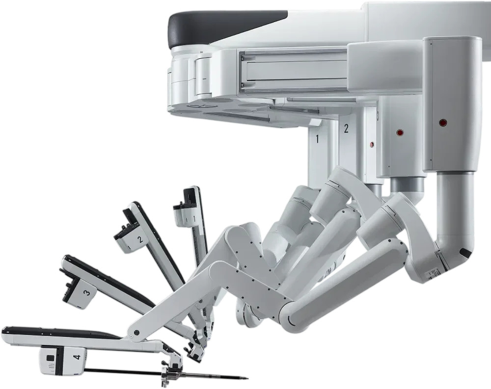
Robotic platforms provide enhanced three-dimensional visualization, refined instrument articulation, and improved precision within confined anatomical spaces. These capabilities are particularly valuable in technically demanding operations such as robotic pancreatic surgery, robotic liver surgery, and complex minimally invasive gastrointestinal cancer procedures.
For appropriately selected patients, robotic techniques may allow for smaller incisions, meticulous tissue handling, reduced postoperative discomfort, shorter hospital stays, and a more efficient recovery — while maintaining rigorous oncologic standards including complete tumor removal and appropriate lymph node evaluation.
These videos are designed to help patients, families, and referring physicians better understand how robotic surgical oncology techniques are applied in real clinical cases, and why a robotic approach may be recommended as part of an individualized treatment strategy.
Our goal is to integrate advanced surgical technology with disciplined oncologic technique and thoughtful, patient-centered care. Dr. Krampitz is recognized as a Master Surgeon in Robotic Surgery by the Surgical Review Corporation, an independent organization that evaluates surgeon experience, outcomes, and adherence to established quality benchmarks in robotic surgery. This commitment allows patients to move forward with clarity, confidence, and a treatment plan grounded in both innovation and experience.
These cases reflect real operative experiences in robotic hepatopancreatobiliary and gastrointestinal surgical oncology and are presented to support education for patients, trainees, and referring physicians.
For physicians seeking referral information or multidisciplinary collaboration, please visit the For Referring Physicians page.
Selected Robotic Operative Demonstrations
The following cases include robotic pancreatic resection, robotic liver resection, biliary surgery, upper gastrointestinal cancer surgery, and colorectal cancer surgery.
These demonstrations are intended for educational purposes and highlight key operative principles including oncologic dissection, vascular control, intraoperative ultrasound guidance, and minimally invasive reconstruction.
Robotic Hepatopancreatobiliary (HPB) Surgery
Hepatopancreatobiliary (HPB) surgery involves the treatment of complex diseases affecting the pancreas, liver, and bile ducts. These operations are among the most technically demanding procedures in abdominal surgery, often requiring meticulous dissection near major blood vessels and delicate biliary structures.
Robotic surgical platforms can enhance visualization and instrument precision in these confined anatomical spaces. High-definition three-dimensional optics and articulating instruments allow refined movements that support careful tumor dissection, vascular preservation, and precise tissue handling.
In appropriately selected patients, robotic techniques may be applied to procedures such as robotic liver surgery, robotic pancreatic surgery, and complex biliary operations, while maintaining the fundamental oncologic principles of complete tumor removal, appropriate lymph node assessment, and multidisciplinary cancer care.
The videos in this library include selected operative demonstrations from robotic HPB and gastrointestinal cancer surgery, illustrating how advanced minimally invasive techniques are integrated with disciplined surgical oncology practice.
Robotic Pancreatic Surgery
Robotic pancreatic surgery may be used for selected pancreatic tumors, including pancreatic cancer and pancreatic neuroendocrine tumors. Robotic platforms provide improved visualization and articulation in the confined retroperitoneal space surrounding the pancreas.
Borderline-resectable pancreatic adenocarcinoma treated with neoadjuvant chemotherapy followed by robotic subtotal distal pancreatectomy and splenectomy, achieving R0 resection with node-negative disease and discharge on postoperative day three.
A 77-year-old woman with a family history of pancreatic cancer and a personal history of cerebrovascular accidentpresented with a biopsy-proven pancreatic ductal adenocarcinoma located in the distal pancreatic neck/proximal body.
Initial staging demonstrated borderline-resectable disease (Type C). She therefore underwent neoadjuvant systemic therapy with gemcitabine and nab-paclitaxel, completing four cycles with favorable radiographic response and improvement in tumor markers, suggesting adequate disease control and candidacy for surgical resection.
Following multidisciplinary review, the patient proceeded to robotic resection using the da Vinci 5 platform.
The operation consisted of:
Robotic staging and exploration
Robotic subtotal distal pancreatectomy
Splenectomy
Cholecystectomy
The procedure was completed minimally invasively without conversion.
Final pathology demonstrated:
Pancreatic ductal adenocarcinoma
pT2 pN0 disease
0 of 18 lymph nodes involved
Negative resection margins (R0)
The patient's postoperative course was uneventful. She recovered well and was discharged home on postoperative day three.
This case illustrates the application of robotic pancreatic resection following neoadjuvant therapy for borderline-resectable pancreatic cancer, demonstrating the ability to achieve oncologically sound resection with lymph node evaluation and margin-negative surgery while maintaining the benefits of minimally invasive recovery.
Robotic completion total pancreatectomy and splenectomy for biopsy-proven locally recurrent pancreatic ductal adenocarcinoma following prior robotic Whipple and systemic therapy, illustrating advanced minimally invasive hepatopancreatobiliary surgical oncology techniques.
This operative video demonstrates a robotic completion total pancreatectomy and splenectomy performed for locally recurrent pancreatic ductal adenocarcinoma (PDAC) following prior pancreatic resection and systemic therapy.
The patient is a 71-year-old man who initially presented with borderline resectable pancreatic cancer involving the pancreatic head, classified as type A due to tumor–vascular interface and type B due to elevated tumor marker CA 19-9. He completed eight cycles of neoadjuvant FOLFIRINOX, demonstrating a favorable radiographic response.
He subsequently underwent a robotic pancreaticoduodenectomy with vascular reconstruction. Final pathology demonstrated ypT1 ypN0 pancreatic ductal adenocarcinoma with 0 of 19 lymph nodes involved and negative surgical margins. The patient then completed four additional cycles of adjuvant FOLFIRINOX, completing a total of 12 cycles of systemic therapy.
During postoperative surveillance, imaging identified a new mass in the body of the pancreas, which was biopsy-proven locally recurrent pancreatic ductal adenocarcinoma.
The patient underwent robotic staging laparoscopy followed by completion total pancreatectomy and splenectomy. Intraoperative ultrasound (IOUS) was used to evaluate pancreatic and vascular anatomy.
The pathology demonstrated pT2 pN1 moderately differentiated pancreatic ductal adenocarcinoma with 2 of 15 lymph nodes involved and negative surgical margins.
The patient recovered well following surgery and was discharged home on postoperative day four, with pancreatic enzyme replacement therapy (PERT) and insulin therapy initiated for management of endocrine and exocrine insufficiency.
This case illustrates the application of advanced robotic techniques in complex pancreatic reoperative surgery within a multidisciplinary hepatopancreatobiliary oncology program.
Robotic distal pancreatectomy and splenectomy for biopsy-proven non-functional pancreatic neuroendocrine tumor of the pancreatic body and tail demonstrating advanced minimally invasive hepatopancreatobiliary surgical oncology techniques.
This operative video demonstrates a robotic distal pancreatectomy and splenectomy performed for a biopsy-proven pancreatic neuroendocrine tumor (PNET) involving the distal body and tail of the pancreas.
The patient is a 77-year-old woman with a medical history notable for hypertension, hyperlipidemia, osteopenia, and prior stage T1bcN0 grade 1 ER/PR-positive, HER2-nonamplified invasive ductal carcinoma of the breast treated with lumpectomy and endocrine therapy.
She presented with abdominal pain, and diagnostic evaluation revealed a 4 cm non-functional, well-differentiated neuroendocrine tumor of the distal pancreas without radiographic evidence of metastatic disease.
The patient underwent robotic distal pancreatectomy and splenectomy.
Final pathology demonstrated:
pT2 pN1 well-differentiated neuroendocrine tumor
Ki-67 index 1.2% (grade 1)
2 of 13 lymph nodes involved with tumor
Lymphovascular invasion present
Negative surgical margins
The patient recovered well following surgery and was discharged home on postoperative day three with a surgical drain in place, which was removed during the first postoperative clinic visit.
This case demonstrates the use of robotic minimally invasive techniques in pancreatic neuroendocrine tumor surgery, allowing precise dissection and lymphadenectomy while maintaining strict oncologic principles.
Robotic subtotal distal pancreatectomy with splenectomy and lymphadenectomy for resectable pancreatic body carcinoma in a high-risk patient, demonstrating celiac axis dissection and PV–SMV confluence exposure.
This case demonstrates a robotic subtotal distal pancreatectomy with splenectomy and regional lymphadenectomyfor a resectable pancreatic carcinoma centered in the pancreatic body in a medically complex patient.
The patient presented with a 3.5 cm pancreatic body mass diagnosed by endoscopic ultrasound–guided biopsy as pancreatic ductal adenocarcinoma with CA 19-9 of 124 U/mL in the setting of normal bilirubin. Cross-sectional imaging demonstrated no evidence of distant metastatic disease and no involvement of the superior mesenteric artery, celiac axis, common hepatic artery, or PV/SMV confluence, consistent with anatomically resectable disease.
The patient’s clinical course was complicated by end-stage renal disease requiring hemodialysis, poorly controlled diabetes, and limited functional status (ECOG 2–3). Medical oncology evaluation concluded that the patient was not an appropriate candidate for systemic therapy, making a surgery-first strategy the most appropriate curative-intent approach.
Robotic staging laparoscopy confirmed no peritoneal metastatic disease. Intraoperative ultrasound localized a ~4 cm tumor within the pancreatic body. The operation included celiac axis dissection to define hepatic, left gastric, and splenic arterial anatomy, creation of a retropancreatic tunnel at the PV/SMV confluence, and pancreatic transection proximal to the tumor with negative proximal margin on frozen section. The splenic artery was divided at its origin from the celiac axis, and the splenic vein was divided distal to the IMV confluence, allowing completion of an en bloc distal pancreatectomy, splenectomy, and regional lymphadenectomy.
Final pathology demonstrated a 4.2 cm medullary carcinoma of the pancreas with negative margins, metastatic carcinoma in 5 of 27 regional lymph nodes, and intact mismatch repair protein expression (pMMR), corresponding to pT3 pN2 disease (AJCC 8th edition). A background low-grade branch-duct IPMN of gastric phenotype was also identified.
The patient recovered well postoperatively and was discharged home on postoperative day seven.
This operative video highlights robotic techniques for complex pancreatic body tumors, including ultrasound-guided tumor localization, celiac axis dissection, retropancreatic tunneling at the PV/SMV confluence, and oncologic lymphadenectomy, demonstrating the application of minimally invasive surgery within disciplined oncologic principles for pancreatic cancer.
Robotic pancreaticoduodenectomy (Whipple procedure) performed for duodenal neuroendocrine tumor causing gastric outlet obstruction, demonstrating advanced minimally invasive hepatopancreatobiliary surgical oncology techniques.
This operative video demonstrates a robotic pancreaticoduodenectomy (Whipple procedure) performed for a duodenal neuroendocrine tumor causing gastric outlet obstruction in an 83-year-old patient.
Robotic techniques were used to facilitate precise dissection of the pancreatic head, duodenum, and surrounding vascular structures while maintaining oncologic principles of complete tumor resection and regional lymph node evaluation.
The patient recovered well following surgery and was discharged home on postoperative day four.
Final pathology demonstrated a well-differentiated neuroendocrine tumor, grade 2 (pT3 pN1) with 3 of 15 lymph nodes involved and negative surgical margins.
This case illustrates the application of robotic minimally invasive techniques in complex pancreatic surgery within a multidisciplinary hepatopancreatobiliary oncology program.
Robotic pancreaticoduodenectomy with portal vein–superior mesenteric vein patch reconstruction using bovine pericardium for borderline-resectable pancreatic ductal adenocarcinoma following neoadjuvant FOLFIRINOX. The video demonstrates robotic vascular control, venous resection, and patch angioplasty at the PV–SMV confluence.
This case demonstrates robotic vascular reconstruction of the portal vein–superior mesenteric vein (PV–SMV) confluence during a robotic pancreaticoduodenectomy.
The patient is a 53-year-old man with biopsy-proven pancreatic ductal adenocarcinoma of the pancreatic head harboring KRAS G12D and TSC1 R245* mutations. Initial staging demonstrated borderline-resectable disease, classified as:
Type A due to tumor involvement of the portal vein–superior mesenteric vein confluence
Type B due to markedly elevated CA 19-9
He completed four cycles of neoadjuvant FOLFIRINOX, with favorable radiographic response and no evidence of metastatic disease.
The patient subsequently underwent:
Robotic staging laparoscopy
Regional lymphadenectomy for staging
Robotic cholecystectomy
Indocyanine green (ICG) cholangiography
ICG angiography
Robotic pancreaticoduodenectomy (Whipple procedure)
During dissection, tumor involvement of the portal vein–SMV confluence necessitated segmental venous resection with patch reconstruction.
Following en bloc resection of the involved venous segment, the portal vein and superior mesenteric vein were reconstructed using a bovine pericardial patch (V1 repair). The robotic platform facilitated precise vascular control, fine dissection around the mesenteric vasculature, and meticulous intracorporeal vascular suturing.
The video highlights the key technical steps of robotic venous reconstruction, including:
vascular control of the PV–SMV confluence
venotomy creation and preparation
bovine pericardial patch sizing and orientation
robotic vascular suturing for patch angioplasty
restoration of laminar venous flow
The patient recovered uneventfully and was discharged home on postoperative day 4.
Robotic Liver Surgery for Liver Tumors and Liver Metastases
Robotic liver surgery can be applied to selected patients with liver tumors and liver metastases. High-definition three-dimensional visualization and precise instrument articulation support meticulous dissection and ultrasound-guided tumor localization during minimally invasive hepatobiliary procedures.
Biopsy-proven colorectal liver metastasis with a concurrent radiographically suspicious RUQ omental lesion (not biopsied pre-op). Robotic staging laparoscopy, omentectomy, and ultrasound-guided microwave ablation of a segment 5 hepatic metastasis. Final omental pathology: fat necrosis, no carcinoma.
A 73-year-old male with previously resected left-sided colorectal adenocarcinoma (pT4aN2a, MSS) status post low anterior resection and adjuvant therapy developed a biopsy-proven hepatic segment 5 metastasis consistent with colorectal origin. Interval cross-sectional imaging and PET also demonstrated a persistent right upper quadrant omental abnormality with mild hypermetabolism, raising concern for regional peritoneal metastatic disease versus inflammatory change. The omental process was not biopsied preoperatively.
He underwent robotic staging laparoscopy to directly evaluate for carcinomatosis and refine candidacy for local therapy. Intraoperatively, a large right omental mass adherent to the anterior abdominal wall was identified and resected with omentectomy, with no other evidence of peritoneal disease. Intraoperative hepatic ultrasound confirmed the known segment 5 lesion and excluded additional liver metastases. Ultrasound-guided microwave ablation was performed to treat the hepatic metastasis with stereotactic targeting, tract ablation, and careful thermal protection of adjacent bowel.
Final pathology from the omentectomy demonstrated fat necrosis with no carcinoma identified, clarifying the etiology of the right upper quadrant finding and confirming absence of diffuse peritoneal spread. The patient recovered well and was discharged home on postoperative day one.
Robotic hepatic cyst fenestration and partial hepatectomy for symptomatic giant hepatic cysts involving the right hepatic lobe. Intraoperative ultrasound and indocyanine green fluorescence imaging guided safe parenchymal transection and biliary identification. The patient was discharged on postoperative day one.
This case demonstrates robotic hepatic cyst fenestration and partial hepatectomy for symptomatic giant hepatic cysts involving the right lobe of the liver.
The patient is a 59-year-old female with progressively symptomatic large hepatic cysts identified on cross-sectional imaging, the largest measuring approximately 14 cm. She reported increasing abdominal discomfort and mass-effect symptoms. Imaging demonstrated numerous hepatic cysts predominantly involving the right hepatic lobe, consistent with benign biliary cystic disease.
Because of the size of the cysts and progressive symptoms, operative management was recommended.
The patient underwent robotic diagnostic laparoscopy, intraoperative hepatic ultrasound, cyst fenestration, partial hepatectomy, and cholecystectomy with indocyanine green (ICG) cholangiography and angiography.
Intraoperative ultrasound demonstrated extensive replacement of the right hepatic lobe by a conglomerate of large hepatic cysts, with distortion of the normal hepatic vascular anatomy due to mass effect. The cyst burden involved a substantial portion of the right hepatic parenchyma, making simple fenestration alone insufficient, and therefore ultrasound-guided partial hepatectomy was performed to remove the dominant cyst complex and decompress the remaining cystic structures.
The right hepatic lobe was mobilized, and the planned transection plane was defined using intraoperative ultrasound and fluorescence guidance. Hepatic parenchymal transection was performed using a clamp-crush technique with saline-coupled bipolar energy and advanced vessel sealing devices. A small segment V biliary tributary encountered along the transection surface was repaired with fine suture after identification of a minor bile leak.
Concurrent robotic cholecystectomy with indocyanine green cholangiography was performed to define biliary anatomy and treat coexisting gallstone disease.
Hemostasis and biliary integrity were confirmed using ICG fluorescence imaging prior to completion of the operation.
Final pathology demonstrated:
Multiloculated simple biliary cysts measuring up to 12 cm
Background hepatic parenchyma with chronic inflammation, reactive biliary proliferation, and pressure-related fibrosis
No dysplasia, atypia, or malignancy
Gallbladder with cholelithiasis and chronic cholecystitis
The patient recovered well following surgery.
A surgical drain placed along the hepatic transection surface was removed prior to discharge, and the patient was discharged home on postoperative day 1.
This case illustrates the role of robotic minimally invasive liver surgery for the treatment of symptomatic giant hepatic cysts, allowing precise parenchymal transection, ultrasound-guided resection, and enhanced visualization of biliary anatomy while minimizing surgical trauma and facilitating rapid recovery.
Robotic ultrasound-guided microwave ablation and wedge resection of bilobar liver metastases from neuroendocrine tumor with mesenteric lymph node metastasis resection, demonstrating advanced minimally invasive hepatopancreatobiliary (HPB) surgical oncology techniques.
This operative video demonstrates robotic ultrasound-guided microwave ablation of multiple bilobar hepatic metastases from an enteric neuroendocrine tumor, combined with wedge resection of a peripheral liver metastasis and resection of a mesenteric lymph node metastasis.
Intraoperative ultrasound was used to localize lesions and guide precise ablation while preserving uninvolved liver parenchyma. Robotic instrumentation facilitates controlled dissection and accurate probe placement during complex hepatobiliary procedures.
The patient recovered well and was discharged home on postoperative day one.
Robotic left hepatectomy with intraoperative ultrasound, Glissonian inflow control, and indocyanine green angiography for solitary liver metastasis from renal cell carcinoma.
This operative video demonstrates a robotic left hepatectomy performed for a solitary liver metastasis from clear cell renal cell carcinoma.
The patient is an 83-year-old man with a history of Stage IV (pT4 pN0) grade 2 clear cell renal cell carcinoma treated with margin-negative left radical nephrectomy in 2016 without adjuvant therapy.
Surveillance imaging initially identified a 1.3 cm hypodense lesion in the left lateral segment of the liver, which was initially interpreted as likely benign. Over time, serial imaging demonstrated progressive enlargement to approximately 6.6–6.8 cm, raising concern for metastatic disease.
Subsequent diagnostic evaluation included:
MRI abdomen demonstrating a 6.6 cm left hepatic mass
PET/CT without hypermetabolic activity but persistent suspicion for malignancy
Endoscopic ultrasound with fine-needle aspiration, confirming metastatic clear cell renal cell carcinoma
Staging evaluation demonstrated no additional sites of metastatic disease, and the patient was referred for surgical management.
The patient underwent robotic staging laparoscopy followed by robotic partial left hepatectomy.
Intraoperative hepatic ultrasound confirmed a solitary lesion in the left lobe with no additional tumors identified in the liver.
A Glissonian approach to the left hepatic pedicle was performed. Hepatomies were created adjacent to the pedicle, allowing passage of a vessel loop behind the left hepatic pedicle in a Glissonian fashion. After confirming preserved perfusion to the right liver using Doppler ultrasound, the left hepatic pedicle was divided using a robotic vascular stapler.
Indocyanine green (ICG) angiography was used to confirm vascular demarcation of the left hepatic lobe, and the line of transection was marked.
Hepatic parenchymal transection was then performed using energy devices and bipolar dissection with selective ligation of vascular and biliary structures. The left hepatic vein was divided using a robotic vascular stapler, and the specimen was removed in an EndoCatch bag.
The resection bed was inspected and demonstrated excellent hemostasis without bile leak. A surgical drain was placed along the transection line.
The patient recovered well and was discharged home on postoperative day three.
Final pathology demonstrated:
Metastatic clear cell renal cell carcinoma
Tumor size 5.7 cm
Nuclear grade 2
Negative surgical margins
This case illustrates the use of robotic minimally invasive techniques for anatomic liver resection, integrating intraoperative ultrasound, Glissonian inflow control, and fluorescence imaging to facilitate precise hepatobiliary surgery.
Robotic right hepatectomy with intraoperative ultrasound-guided microwave ablation for colorectal cancer liver metastases following systemic chemotherapy, illustrating advanced minimally invasive hepatopancreatobiliary surgical oncology techniques.
This operative video demonstrates a robotic right hepatic lobectomy with ultrasound-guided microwave ablationperformed for colorectal cancer liver metastases following systemic chemotherapy.
The patient is a 49-year-old woman with biopsy-proven microsatellite stable (MSS) sigmoid adenocarcinomainitially staged MRI T3N0 with PET/CT evidence of metastatic disease (M1) involving three hepatic lesions located in segments 2/4A, 5/6, and 8.
She completed 12 cycles of modified FOLFOX (mFOLFOX), achieving favorable systemic disease control. Restaging PET/CT demonstrated persistent metabolic activity limited to the segment 5/6 hepatic lesion and the rectosigmoid primary, with no evidence of additional metastatic disease.
Following multidisciplinary review and favorable restaging imaging, the patient underwent a staged minimally invasive surgical approach including:
Robotic low anterior resection for sigmoid colon cancer
Robotic staging laparoscopy
Intraoperative hepatic ultrasound
Ultrasound-guided microwave ablation of the segment 2/4A lesion
Indocyanine green (ICG) cholangiography
Robotic cholecystectomy
Robotic right hepatic lobectomy
This approach allowed treatment of multifocal colorectal liver metastases using a combination of anatomic resection and targeted ablation, while preserving uninvolved hepatic parenchyma.
The patient recovered well following surgery and was discharged home on postoperative day three.
This case illustrates the integration of systemic therapy, advanced imaging, and robotic hepatobiliary surgery in the multidisciplinary management of colorectal liver metastases.
Learn more about Colon Cancer and Colorectal Liver Metastases.
Robotic Biliary Surgery
Robotic techniques can also be applied to complex biliary procedures involving the bile ducts and gallbladder, including operations for cholangiocarcinoma and other hepatobiliary conditions requiring precise dissection near critical vascular structures.
Robotic completion hepatic lymphadenectomy for pT2 gallbladder adenocarcinoma following prior robotic cholecystectomy and segment IVb/V resection.
This operative video demonstrates a robotic completion hepatic lymphadenectomy performed for pT2 gallbladder adenocarcinoma following prior radical cholecystectomy.
The patient is a 73-year-old woman with a history of diabetes mellitus, hyperlipidemia, and prior breast cancer who was incidentally found to have gallbladder wall thickening on imaging performed during evaluation for an unrelated condition.
Subsequent evaluation demonstrated hypermetabolic gallbladder wall thickening on PET/CT, concerning for malignancy without evidence of distant metastatic disease.
The patient underwent a robotic cholecystectomy with segment IVb/V hepatic resection and portal lymphatic dissection performed by another surgeon. Final pathology demonstrated:
Biliary-type moderately differentiated adenocarcinoma
pT2a disease (tumor invading perimuscular connective tissue on the peritoneal side)
Negative surgical margins
Perineural invasion present
No regional lymph nodes identified in the specimen (pNx)
Postoperative imaging and tumor markers did not demonstrate evidence of residual or metastatic disease.
Because adequate regional lymph node staging had not been performed, the case was reviewed at a multidisciplinary tumor board. Current oncologic guidelines recommend regional lymphadenectomy for pT2 gallbladder cancer, given the relatively high incidence of nodal metastasis and the prognostic importance of nodal status.
The patient was therefore offered completion hepatic lymphadenectomy for staging and potential therapeutic benefit.
The patient underwent robotic staging laparoscopy and completion lymphadenectomy, including dissection of lymphatic tissue along the:
Common bile duct
Porta hepatis
Common hepatic and left hepatic arteries
Celiac axis
Retropancreatic lymph node basin
Intraoperative findings included postoperative adhesions without evidence of metastatic disease.
The patient tolerated the procedure well and was discharged home on postoperative day one.
Final pathology demonstrated:
No metastatic carcinoma identified in six regional lymph nodes (0/10)
This case illustrates the role of completion lymphadenectomy in pT2 gallbladder cancer when adequate nodal staging was not performed at the time of the initial radical cholecystectomy.
A 49-year-old patient with a history of laparoscopic cholecystectomy presented with recurrent cholangitis and persistent biliary symptoms despite multiple endoscopic interventions. Prior ERCP procedures demonstrated recurrent common bile duct stones and a large retained cystic duct remnant containing stones, which was not amenable to endoscopic clearance due to the narrow and tortuous cystic duct–common bile duct junction.
Cross-sectional imaging and endoscopic studies demonstrated a markedly dilated cystic duct remnant measuring approximately 3–4 cm with a large retained stone, functioning as a reservoir for recurrent biliary stasis and stone formation. This anatomy is a recognized cause of post-cholecystectomy syndrome, particularly when the cystic duct stump is long or when surgical clips are placed proximally.
The patient underwent robotic diagnostic laparoscopy, extensive adhesiolysis, intraoperative ultrasound, indocyanine green cholangiography, cystic duct remnant resection, and common bile duct exploration.
Intraoperative ultrasound confirmed a dilated cystic duct remnant communicating with the common bile duct and containing a large stone. After careful dissection of the hepatoduodenal ligament, the cystic duct remnant was opened and the stone extracted. The remnant was then divided flush with the common bile duct using a robotic vascular stapler to avoid ductal narrowing. Indocyanine green cholangiography confirmed biliary continuity and absence of bile leak.
A falciform ligament pedicle flap was used to buttress the transection site.
The patient recovered uneventfully and was discharged following an uncomplicated postoperative course.
This case illustrates the role of robotic hepatobiliary surgery in the management of complex post-cholecystectomy biliary pathology, particularly when endoscopic approaches are unsuccessful.
Robotic outpatient cholecystectomy demonstrating safe dissection of the hepatocystic triangle and critical view of safety for symptomatic gallstones.
This case demonstrates robotic cholecystectomy for symptomatic cholelithiasis in a 48-year-old female patient with obesity (BMI 36) and a three-year history of progressive intermittent post-prandial right upper quadrant pain.
Diagnostic evaluation included abdominal ultrasound, which demonstrated cholelithiasis without evidence of acute cholecystitis or biliary obstruction, consistent with symptomatic gallstone disease (biliary colic).
Symptomatic cholelithiasis is a common condition, and minimally invasive cholecystectomy is one of the most frequently performed outpatient operations in general surgery. When symptoms develop, elective cholecystectomy is recommended because gallstone disease may progress to more serious complications, including acute cholecystitis, choledocholithiasis, gallstone pancreatitis, or cholangitis, which can increase both morbidity and procedural complexity.
The operation was performed using a robotic minimally invasive approach, which provides enhanced visualization and instrument articulation during dissection of the hepatocystic triangle.
Key steps demonstrated in this video include:
Exposure of the hepatocystic triangle
Identification of the cystic duct and cystic artery
Achievement of the critical view of safety
Secure ligation and division of the cystic structures
Gallbladder removal from the hepatic bed
Robotic cholecystectomy is a routine outpatient operation that allows safe treatment of symptomatic gallstone disease while facilitating rapid recovery.
Robotic Upper Gastrointestinal Cancer Surgery
Robotic surgical platforms can enhance visualization and precision during operations involving the stomach, esophagus, small intestine, colon, and other complex abdominal organs. These procedures often require meticulous dissection in confined anatomical spaces, careful preservation of vascular structures, and precise reconstruction.
In appropriately selected patients, robotic techniques may be applied to gastric cancer surgery, esophageal surgery, colorectal cancer surgery, neuroendocrine tumor resection, and other complex abdominal oncologic procedures. High-definition three-dimensional visualization and articulating instruments can facilitate refined dissection and reconstruction while maintaining strict oncologic principles.
The following operative videos demonstrate selected cases of robotic upper gastrointestinal and complex abdominal oncologic surgery, illustrating how minimally invasive techniques are integrated with disciplined surgical oncology practice.
Robotic Ivor Lewis esophagectomy for gastroesophageal junction adenocarcinoma following neoadjuvant FLOT chemotherapy with durvalumab, demonstrating minimally invasive conduit creation, thoracic lymphadenectomy, and robotic hand-sewn esophagogastric anastomosis.
Robotic Ivor Lewis esophagectomy with lymphadenectomy and two-layer hand-sewn thoracic esophagogastrostomy for locally advanced Siewert type II/III gastroesophageal junction adenocarcinoma following neoadjuvant FLOT chemotherapy with durvalumab.
Pre-treatment staging laparoscopy with peritoneal washings confirmed the absence of occult metastatic disease, allowing the patient to proceed with perioperative systemic therapy based on the MATTERHORN trial regimen. Molecular profiling demonstrated microsatellite-stable disease with low tumor mutational burden (4 muts/Mb) and genomic alterations including CCND1, FGF19, FGF3, FGF4, FLT3, KRAS, and MYC amplifications with TP53 mutation (R282W) and no ERBB2 amplification.
Restaging evaluation demonstrated a favorable treatment response with reduction in tumor size on cross-sectional imaging and marked biochemical response, with CEA decreasing from 57.7 ng/mL to 4.1 ng/mL during neoadjuvant therapy.
The patient subsequently underwent robotic staging laparoscopy, lymphadenectomy, and minimally invasive Ivor Lewis esophagectomy. The operation included mobilization of the stomach with preservation of the right gastroepiploic arcade, division of the left gastric vessels, creation of a narrow gastric conduit, and mediastinal esophageal dissection with paraesophageal and subcarinal lymphadenectomy. A two-layer robotic hand-sewn esophagogastric anastomosis was constructed within the thoracic cavity following conduit perfusion assessment using indocyanine green fluorescence angiography.
Final pathology demonstrated residual moderately differentiated adenocarcinoma of the gastroesophageal junction measuring 2.2 cm with near-complete treatment response, negative margins, and metastatic carcinoma in 4 of 24 lymph nodes (ypT3N2).
The patient recovered without major complications, demonstrated no anastomotic leak on postoperative contrast esophagram, and was discharged home on postoperative day four on a full liquid diet. Systemic therapy was reinitiated five weeks following surgery as part of the perioperative treatment strategy.
Robotic partial gastrectomy for an enlarging gastric gastrointestinal stromal tumor with intraoperative endoscopy and leak testing.
This operative video demonstrates a robotic partial gastrectomy performed for a gastrointestinal stromal tumor (GIST) of the gastric fundus.
The patient is a 59-year-old woman who presented with an enlarging gastric mass consistent with a gastrointestinal stromal tumor on prior biopsy.
The patient underwent robotic staging laparoscopy followed by robotic partial gastrectomy.
Intraoperative evaluation demonstrated a 2.5 cm exophytic lesion arising from the anterior aspect of the gastric fundus. The tumor was elevated and resected using a robotic stapled wedge resection.
Frozen section analysis demonstrated a close margin at the staple line, and therefore the staple line was re-excised to obtain an additional margin. The gastrotomy was then closed transversely with a running hand-sewn suture.
An intraoperative upper endoscopy was performed to evaluate the gastric lumen and the repair. The stomach was insufflated while the repair site was submerged intra-abdominally, and a hydropneumatic leak test confirmed the integrity of the closure.
The patient tolerated the procedure well and was discharged home on postoperative day two.
Final pathology demonstrated:
Gastrointestinal stromal tumor, spindle cell type
Tumor size 4.5 cm
Mitotic rate <5 per 5 mm²
Very low risk category
All margins negative for tumor
This case demonstrates the application of robotic minimally invasive techniques for gastric GIST resection, including precise tumor localization, margin assessment, and intraoperative endoscopic evaluation.
Robotic splenectomy with intraoperative ultrasound for FDG-avid splenic lesions suspicious for lymphoma in a patient with prior splenic artery embolization.
This operative video demonstrates a robotic splenectomy with intraoperative ultrasound performed for FDG-avid splenic lesions suspicious for hematologic malignancy.
The patient is a 64-year-old woman with a complex medical history, including thalassemia, insulin-dependent diabetes mellitus, chronic kidney disease, antiphospholipid syndrome requiring chronic anticoagulation, and idiopathic thrombocytopenic purpura. She previously experienced hemorrhagic shock due to splenic hemorrhage, which was treated with transarterial splenic artery coil embolization.
The patient later presented with sepsis, hypercalcemia, and enlarging FDG-avid splenic lesions, raising strong suspicion for an underlying hematologic malignancy versus infectious process. Prior bone marrow biopsies were nondiagnostic, and percutaneous biopsy of the splenic lesions was considered prohibitively high risk due to the patient’s complex comorbidities and prior splenic artery embolization.
After multidisciplinary evaluation, the patient was offered diagnostic and therapeutic splenectomy.
The patient underwent robotic staging laparoscopy followed by robotic splenectomy with intraoperative ultrasound.
Intraoperative findings included:
Inflammatory adhesions
Splenomegaly with a large splenic mass
Evidence of prior splenic artery coil embolization
The spleen was mobilized by dividing the gastrocolic ligament and short gastric vessels, allowing entry into the lesser sac and exposure of the splenic hilum. The splenocolic ligament was divided and the splenic flexure mobilized to facilitate complete splenic mobilization.
Careful dissection was performed to identify and preserve the tail of the pancreas, which was separated from the splenic hilar vessels prior to vascular division.
The splenic artery and splenic vein were divided using a robotic vascular stapler, and the specimen was retrieved in an EndoCatch bag through a Pfannenstiel extraction incision.
The patient tolerated the procedure well without intraoperative complications.
Final pathology demonstrated:
Diffuse large B-cell lymphoma involving the spleen
High proliferative index (Ki-67 >90%)
Fluorescence in situ hybridization analysis did not demonstrate MYC, BCL2, or BCL6 rearrangements, excluding high-grade double-hit lymphoma.
This case illustrates the role of robotic splenectomy in the diagnosis and management of suspected splenic lymphoma, particularly in medically complex patients in whom bone marrow biopsy is nondiagnostic and percutaneous splenic biopsy is unsafe.
Robotic staging laparoscopy and feeding jejunostomy placement for metastatic gastroesophageal junction adenocarcinoma causing severe dysphagia and malnutrition.
This case demonstrates robotic staging laparoscopy and feeding jejunostomy placement for nutritional support in a patient with gastroesophageal junction (GEJ) adenocarcinoma causing progressive dysphagia and cancer-associated malnutrition.
The patient presented with grade 3 dysphagia and weight loss and was found to have biopsy-proven GEJ adenocarcinoma characterized by microsatellite stability (MSS) and PD-L1 CPS 3, with HER2 status pending at the time of evaluation. Cross-sectional imaging and PET/CT demonstrated a hypermetabolic GEJ mass with thoracic, abdominal, and retroperitoneal lymphadenopathy, as well as indeterminate pulmonary nodules suspicious for metastatic disease.
Evaluation also identified a synchronous descending colon adenocarcinoma, further complicating oncologic management.
Because of the patient’s multiple comorbidities, frailty, and marginal performance status (ECOG 2–3), definitive surgical resection was not appropriate. Multidisciplinary evaluation recommended systemic therapy and/or definitive chemoradiation. However, the patient’s progressive dysphagia and declining nutritional status required long-term enteral access prior to treatment.
A robotic staging laparoscopy was performed to evaluate for occult peritoneal metastases. No intra-abdominal metastatic disease was identified. A feeding jejunostomy tube was then placed robotically by securing a loop of proximal jejunum to the abdominal wall and inserting a 16-French jejunostomy tube using a purse-string and circumferential fixation technique.
The patient recovered well and was discharged home the same day.
Feeding jejunostomy placement is a commonly performed procedure in patients with esophageal and gastroesophageal junction cancers who develop significant dysphagia, allowing reliable enteral nutrition during systemic therapy or chemoradiation.
This video demonstrates robotic techniques for laparoscopic staging and jejunostomy placement in the multidisciplinary management of advanced upper gastrointestinal malignancies.
Robotic Ivor Lewis esophagectomy with lymphadenectomy and hand-sewn esophagogastric anastomosis for distal esophageal neuroendocrine carcinoma following neoadjuvant chemoradiation.
This operative video demonstrates a robotic Ivor Lewis esophagectomy performed for poorly differentiated small cell neuroendocrine carcinoma of the distal esophagus following neoadjuvant chemoradiation.
The patient is a 65-year-old man with a history of hypertension, chronic obstructive pulmonary disease, prior tobacco use, and alcohol exposure who presented with progressive dysphagia, post-prandial discomfort, and a 20-pound weight loss.
Upper endoscopy revealed a large ulcerating mass located 32 cm from the incisors, involving the distal esophagus and gastroesophageal junction. Biopsy demonstrated poorly differentiated small cell neuroendocrine carcinoma, microsatellite stable, with genomic alterations including NOTCH1Q475fs*156, RB1 loss, and TP53 mutation.
Staging FDG-PET/CT demonstrated a 7 cm hypermetabolic distal esophageal mass extending into the gastroesophageal junction, with a prominent perigastric lymph node but no definite evidence of distant metastatic disease.
The patient underwent multimodality therapy, including:
Definitive chemoradiation
Cisplatin and etoposide chemotherapy
Radiation therapy (50.7 Gy in 27 fractions)
Restaging PET/CT demonstrated marked metabolic and radiographic response without progression of disease.
Following multidisciplinary evaluation, the patient underwent robotic staging laparoscopy followed by robotic Ivor Lewis esophagectomy.
The abdominal phase included:
Gastric mobilization and conduit creation
Division of the left gastric artery with lymphadenectomy
Botulinum toxin pyloroplasty
Creation of a 3–4 cm gastric conduit
The thoracic phase included:
Robotic mediastinal esophageal mobilization
Division of the azygos vein
Paraesophageal and subcarinal lymphadenectomy
Construction of a two-layer robotic hand-sewn intrathoracic esophagogastric anastomosis
The patient recovered well following surgery and was discharged home on postoperative day four on a full liquid diet, which was advanced to a regular diet at the first postoperative clinic visit the following week.
Final pathology demonstrated complete pathologic response to neoadjuvant therapy:
No residual viable neuroendocrine carcinoma
0 of 21 lymph nodes involved with tumor
All surgical margins negative
These findings correspond to ypT0 disease with complete treatment response.
This case illustrates the application of robotic minimally invasive esophagectomy following multimodality therapy, demonstrating advanced thoracic and upper gastrointestinal oncologic surgery.
Robotic small bowel resection with mesenteric lymphadenectomy and intracorporeal enteroenterostomy for metastatic ileal neuroendocrine tumor demonstrating minimally invasive resection of the primary tumor and nodal disease.
Robotic small bowel resection with mesenteric lymphadenectomy and intracorporeal enteroenterostomy for well-differentiated ileal neuroendocrine tumor with regional nodal metastases and diffuse hepatic metastatic disease.
The patient presented with a grade 1 (Ki-67 2%) well-differentiated neuroendocrine tumor of the terminal ileumwith mesenteric nodal metastases and numerous small hepatic metastases identified on cross-sectional imaging and DOTATATE PET imaging. The patient had no clinical or biochemical evidence of carcinoid syndrome, although chromogranin A was elevated at 524. Genetic evaluation was performed due to family history suggestive of a hereditary syndrome, but no actionable germline mutations were identified.
Because the diffuse hepatic metastases were not amenable to surgical cytoreduction or liver-directed therapy, operative management focused on resection of the primary tumor and regional nodal disease to reduce the long-term risk of small bowel obstruction, mesenteric ischemia, bleeding, or perforation—well-recognized complications of untreated small bowel neuroendocrine tumors.
Robotic staging laparoscopy confirmed the absence of peritoneal carcinomatosis. The terminal ileal primary tumor and associated mesenteric nodal disease were resected en bloc with regional mesenteric lymphadenectomy. Indocyanine green fluorescence angiography was used to confirm adequate bowel perfusion prior to creation of a robotic intracorporeal stapled enteroenterostomy.
Final pathology demonstrated a 1.8 cm well-differentiated grade 1 ileal neuroendocrine tumor invading through the serosa (pT4) with metastatic tumor in 4 of 4 regional lymph nodes (pN1) and negative surgical margins.
The patient recovered without complications and was discharged home on postoperative day three.
This video demonstrates robotic surgical management of metastatic small bowel neuroendocrine tumor, highlighting techniques for mesenteric nodal dissection near the superior mesenteric vessels and minimally invasive bowel reconstruction.
Robotic staging laparoscopy and celiac axis lymphadenectomy for oligoprogressive metastatic esophageal squamous cell carcinoma following systemic therapy.
This operative video demonstrates a robotic staging laparoscopy and celiac axis lymphadenectomy performed for oligoprogressive metastatic esophageal squamous cell carcinoma.
The patient is a 61-year-old man with HER2-negative, PD-L1 CPS 2 moderately differentiated squamous cell carcinoma of the mid esophagus who initially underwent definitive chemoradiation with weekly carboplatin and paclitaxel.
During treatment the patient developed severe dysphagia and inability to tolerate oral intake, requiring placement of a robotic feeding jejunostomy with subsequent revision for obstruction.
Restaging imaging following chemoradiation unfortunately demonstrated metastatic disease, and the patient was not a candidate for curative esophagectomy. He underwent palliative esophageal stenting and continued systemic therapy with an overall mixed response.
Over time the patient experienced substantial tumor regression and improvement in symptoms, allowing removal of his feeding jejunostomy. However, surveillance imaging demonstrated persistent progression within gastroesophageal and celiac axis lymph nodes while the remainder of disease remained controlled.
Following multidisciplinary discussion, the patient was referred for surgical resection of this limited site of progressionas a palliative disease-control strategy.
The patient underwent robotic staging laparoscopy and celiac axis lymphadenectomy.
Intraoperative findings demonstrated:
Bulky lymph nodes along the left gastric artery and celiac axis
No evidence of additional intra-abdominal metastatic disease
The pars flaccida was opened, allowing exposure of the left gastric artery and celiac axis. The left gastric artery and vein were ligated, and the celiac lymph node packet was dissected from the splenic artery, lesser curvature of the stomach, gastroesophageal junction, diaphragmatic crura, and retroperitoneum.
The specimen was removed in an EndoCatch retrieval bag.
The patient tolerated the procedure well and was discharged home the following day.
Final pathology demonstrated:
Metastatic keratinizing squamous cell carcinoma
1 of 3 celiac lymph nodes involved with tumor
This case illustrates the use of robotic minimally invasive lymphadenectomy for oligoprogressive metastatic disease, performed as a palliative intervention aimed at achieving regional disease control during ongoing systemic therapy.
Robotic Colorectal Cancer Surgery
Robotic platforms can enhance visualization and precision during surgery for colon and rectal cancers, particularly in anatomically confined regions such as the deep pelvis. High-definition three-dimensional optics and articulating instruments allow refined dissection around critical vascular and autonomic structures while supporting meticulous oncologic technique.
In appropriately selected patients, robotic techniques may be used for procedures such as right hemicolectomy, sigmoid colectomy, and low anterior resection. These approaches allow precise vascular ligation, oncologic lymphadenectomy, and intracorporeal reconstruction while maintaining established principles of colorectal cancer surgery.
The following operative videos demonstrate selected cases of robotic colorectal cancer surgery, illustrating how minimally invasive techniques are integrated with disciplined surgical oncology practice.
Robotic low anterior resection with stapled colorectal anastomosis for rectosigmoid adenocarcinoma demonstrating advanced minimally invasive colorectal cancer surgery techniques.
This operative video demonstrates a robotic low anterior resection with stapled colorectal anastomosis performed for rectosigmoid adenocarcinoma.
The patient is an 84-year-old woman, a retired psychiatrist, with a history of bilateral invasive ductal carcinoma of the breast treated with margin-negative bilateral modified radical mastectomy and sentinel lymph node biopsy on November 6, 2024. Her breast cancers were hormone receptor–positive (ER-positive), HER2-negative tumors, and she is currently receiving endocrine therapy with anastrozole.
She was subsequently diagnosed with pMMR well-differentiated adenocarcinoma of the rectosigmoid colon.
The patient underwent robotic staging laparoscopy followed by robotic low anterior resection, including:
Stapled end-to-end colorectal anastomosis
Hydropneumatic leak testing
Flexible sigmoidoscopy for anastomotic assessment
Final pathology demonstrated:
pT1 pN0 moderately differentiated adenocarcinoma
0 of 50 lymph nodes involved with carcinoma
The patient recovered well following surgery and was discharged home on postoperative day three.
This case illustrates the application of robotic minimally invasive colorectal surgery for early-stage rectosigmoid cancer, demonstrating precise pelvic dissection and oncologic lymphadenectomy with safe intracorporeal reconstruction.
Robotic right hemicolectomy with intracorporeal side-to-side enterocolic anastomosis and indocyanine green angiography for nearly obstructing cecal adenocarcinoma demonstrating advanced minimally invasive colorectal cancer surgery techniques.
This operative video demonstrates a robotic right hemicolectomy with intracorporeal enterocolic anastomosisperformed for cecal adenocarcinoma.
The patient is a 95-year-old woman who presented with symptoms of bowel obstruction and was found on colonoscopy to have a nearly obstructing cecal mass. Endoscopic biopsy was non-diagnostic. Staging evaluation demonstrated a CEA of 3.1 with no radiographic evidence of distant metastatic disease.
The patient underwent robotic staging laparoscopy followed by robotic right hemicolectomy. Intraoperative indocyanine green (ICG) angiography was used to assess perfusion of the bowel prior to reconstruction. An intracorporeal side-to-side enterocolic anastomosis was performed using minimally invasive techniques.
Final pathology demonstrated:
pT3 pN2b poorly differentiated adenocarcinoma
8 of 14 lymph nodes involved with carcinoma
Negative surgical margins
The patient recovered well following surgery and was discharged home on postoperative day four.
This case illustrates the application of robotic minimally invasive colorectal surgery in elderly patients with obstructing colon cancer, demonstrating safe oncologic resection and intracorporeal reconstruction.
Robotic Surgical Oncology in the San Francisco Bay Area
Dr. Geoffrey W. Krampitz is a fellowship-trained robotic surgical oncologist specializing in pancreatic cancer surgery, liver tumor surgery, bile duct cancer surgery, and complex gastrointestinal cancers. His practice focuses on advanced minimally invasive and robotic surgery for hepatopancreatobiliary (HPB) and gastrointestinal malignancies in the San Francisco Bay Area.
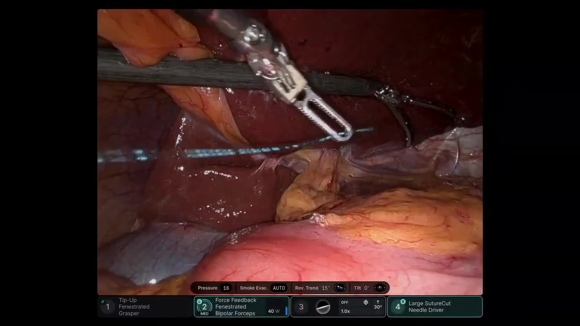
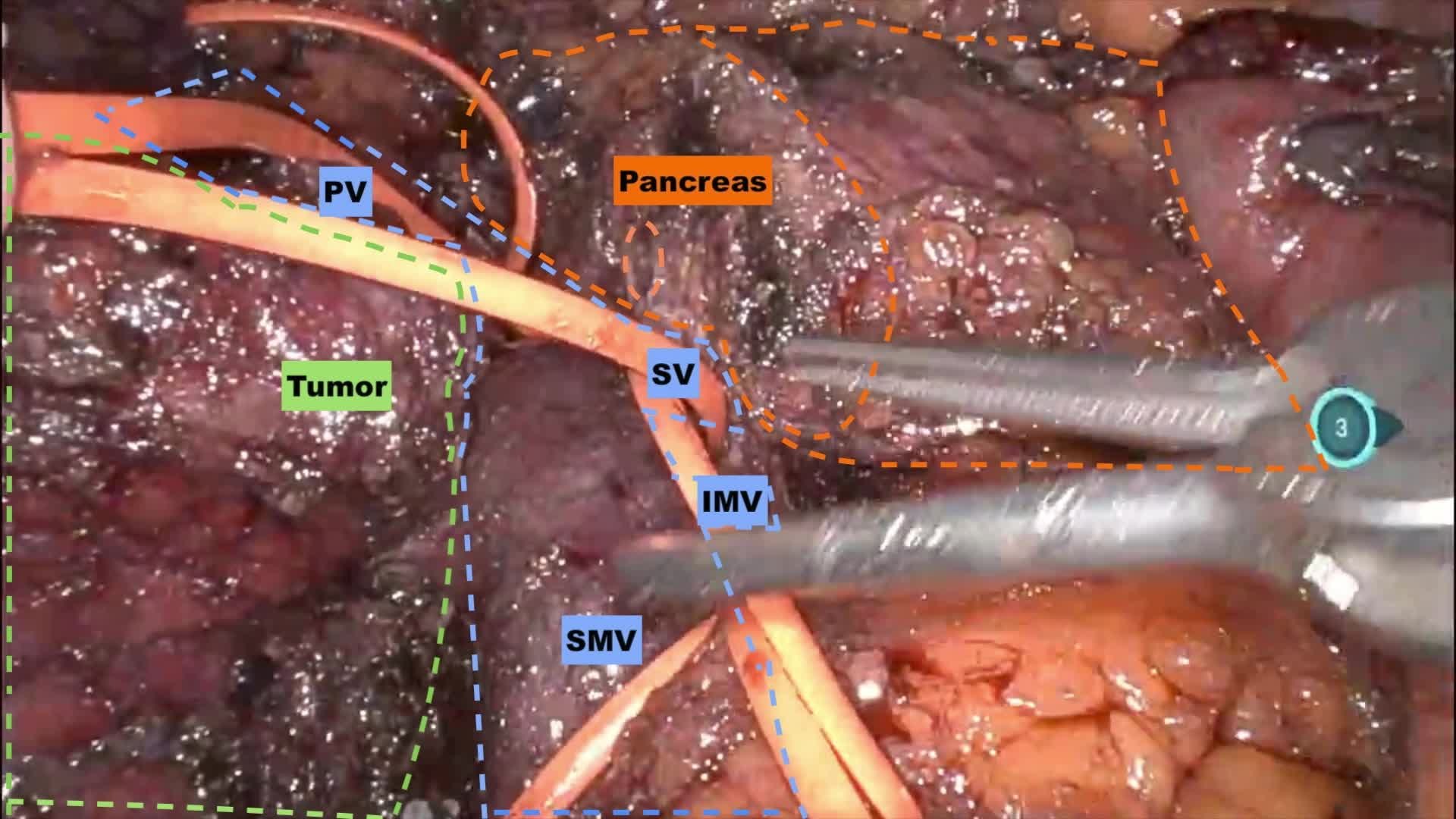
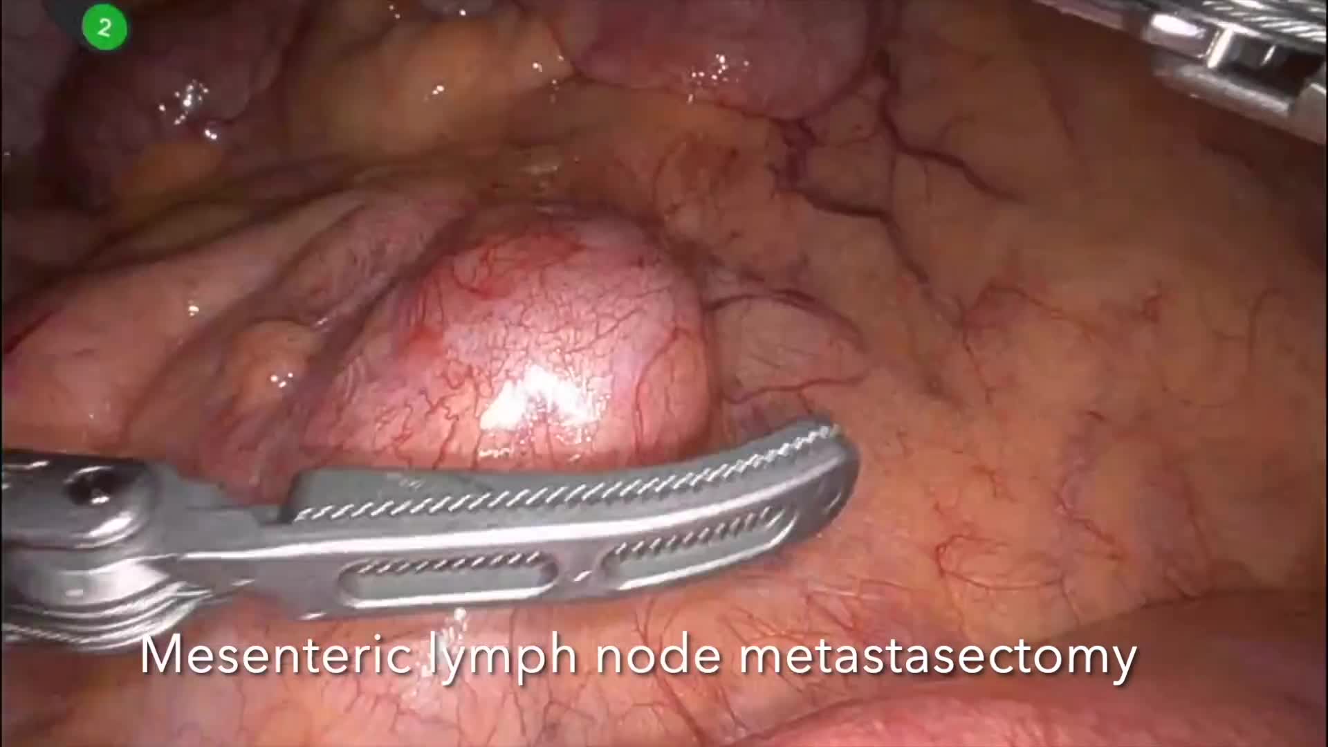
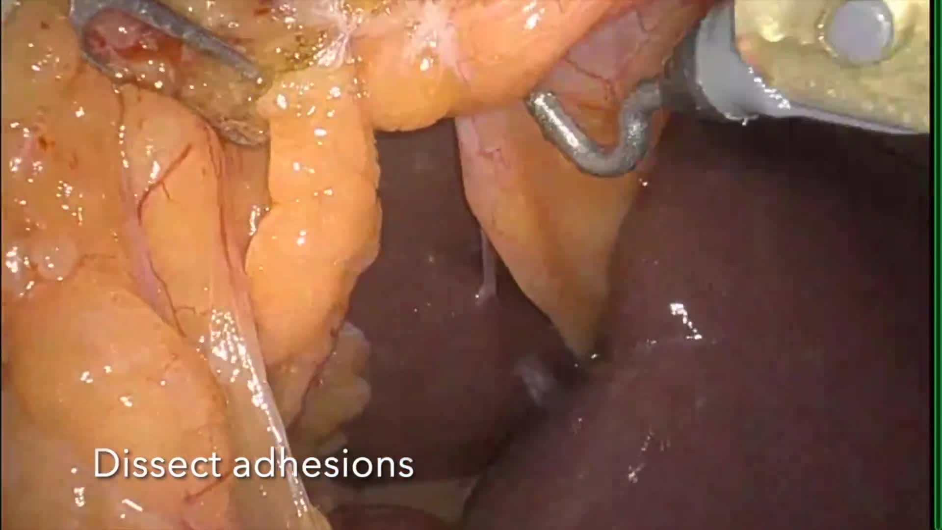
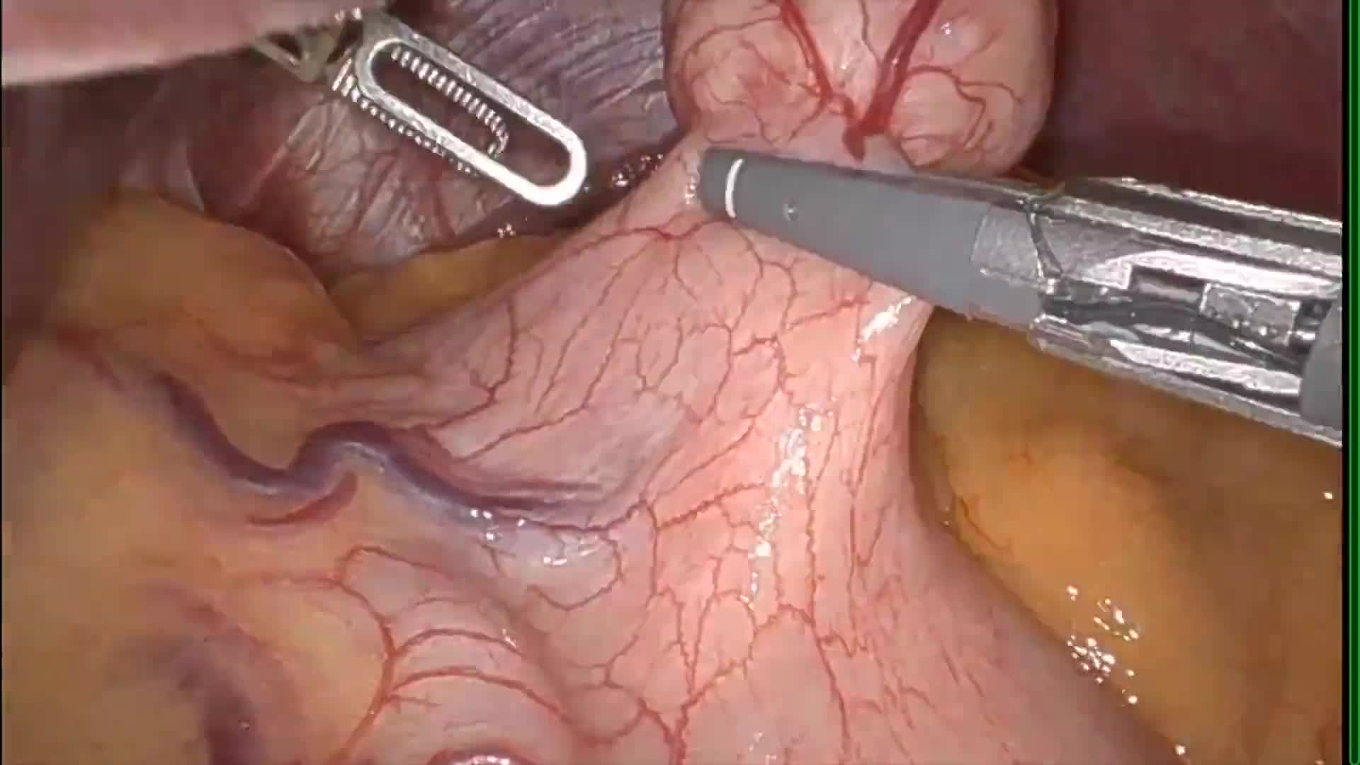
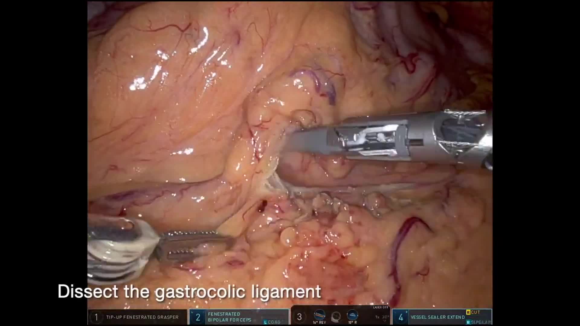
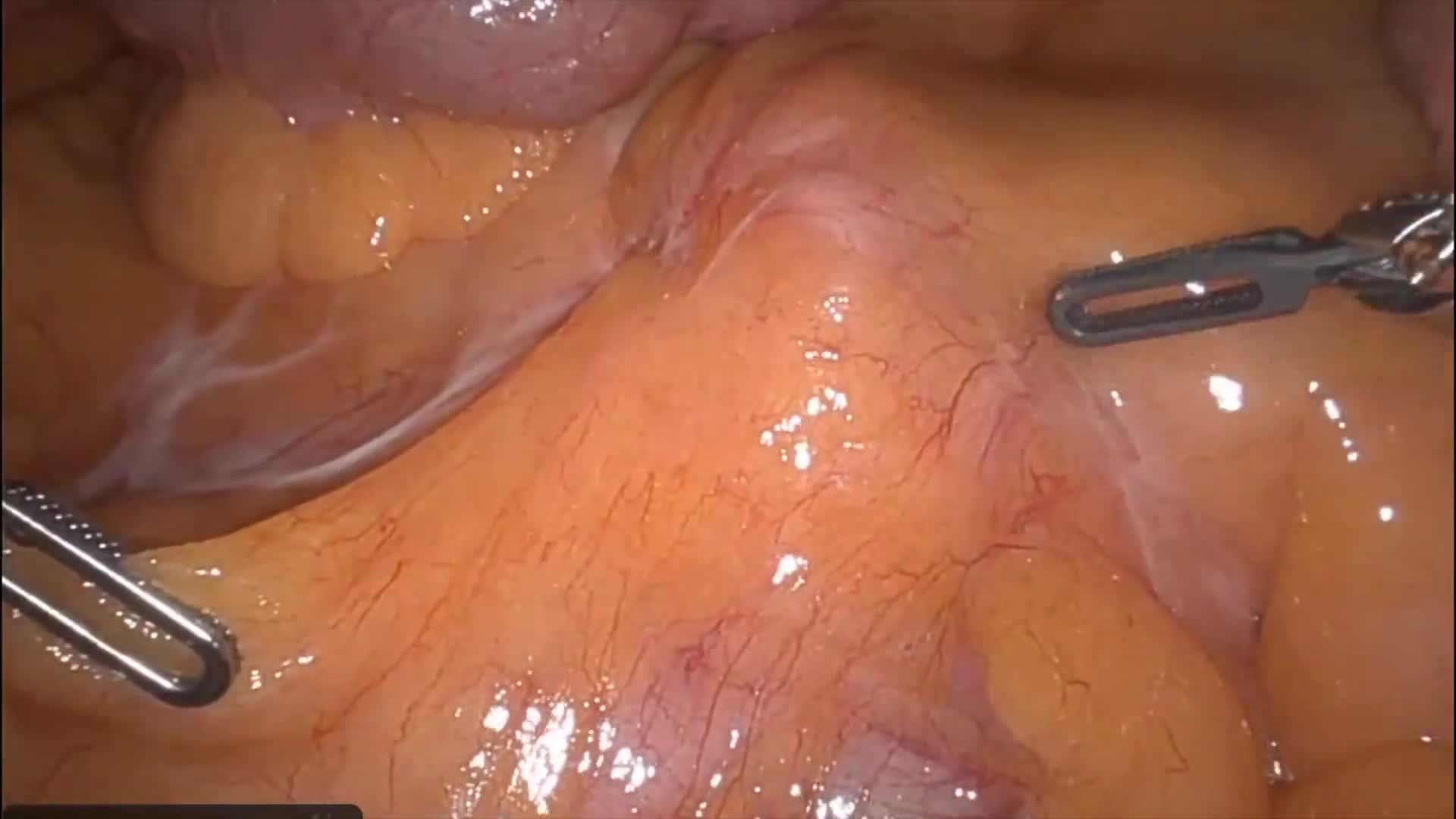
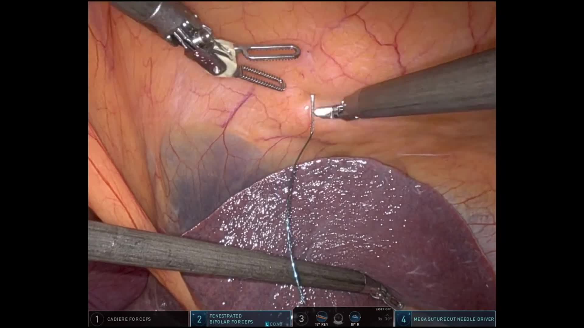
Robotic enucleation of a pancreatic neuroendocrine tumor with lymphadenectomy after prior ileal NET resection, demonstrating ultrasound-guided tumor localization and suprapancreatic dissection.
This case demonstrates robotic enucleation of a pancreatic neuroendocrine tumor with regional lymphadenectomyin a patient previously treated for an ileal neuroendocrine tumor.
The patient initially presented with small bowel obstruction, and workup revealed a 1.7 cm well-differentiated ileal neuroendocrine tumor (WHO grade 1, Ki-67 <3%). He underwent laparoscopic small bowel resection, with pathology demonstrating pT3 pN1 disease with two of three regional lymph nodes involved.
Subsequent staging with DOTATATE PET/CT demonstrated intense radiotracer uptake in two nodular structures posterior to the pancreatic neck within the peripancreatic/gastrohepatic ligament region. Although initially interpreted as possible nonregional nodal metastases, the imaging characteristics and anatomic location also raised the possibility of a primary intrapancreatic neuroendocrine tumor.
Given the limited distribution of disease and the diagnostic uncertainty, surgical exploration was recommended.
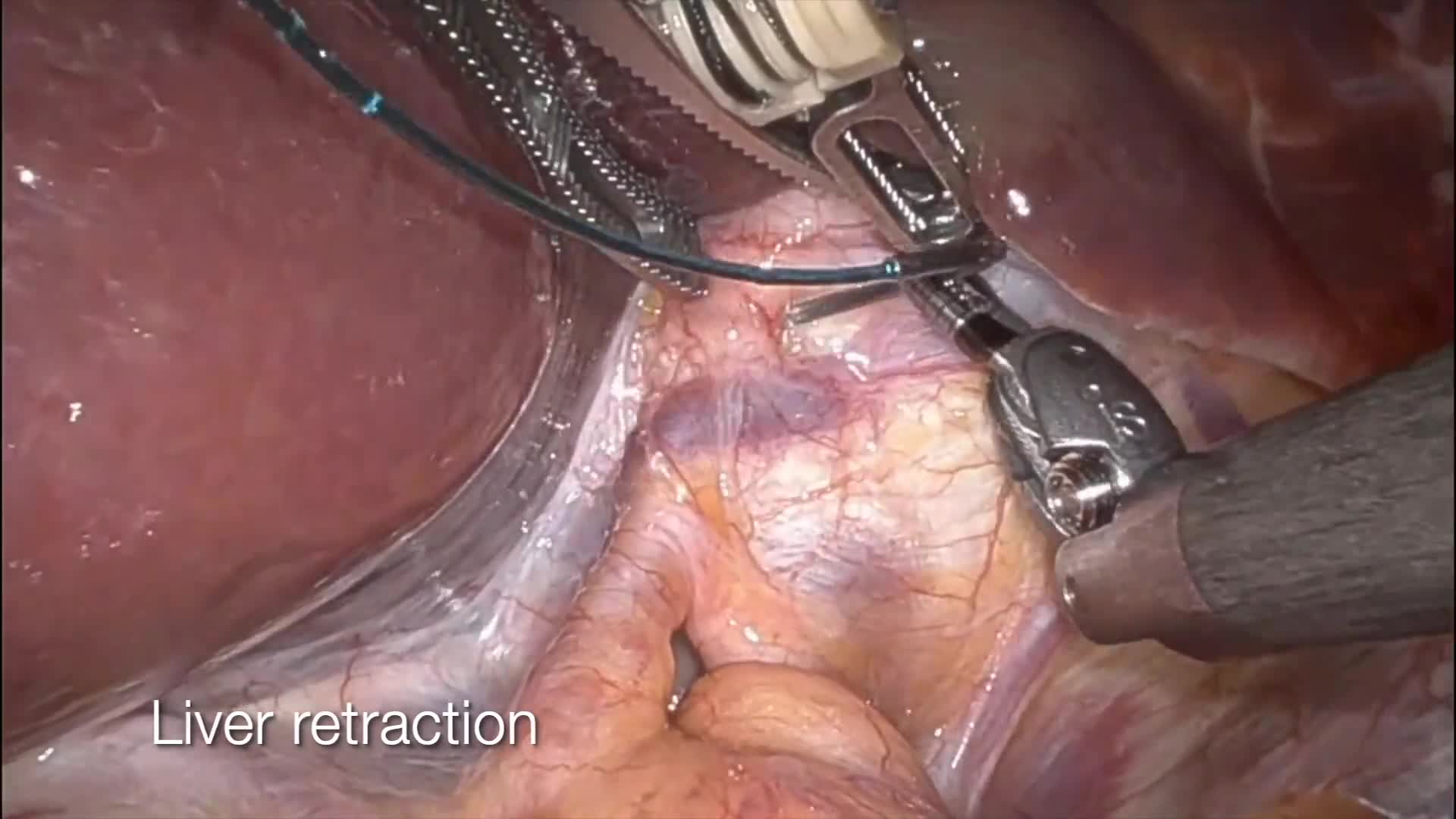
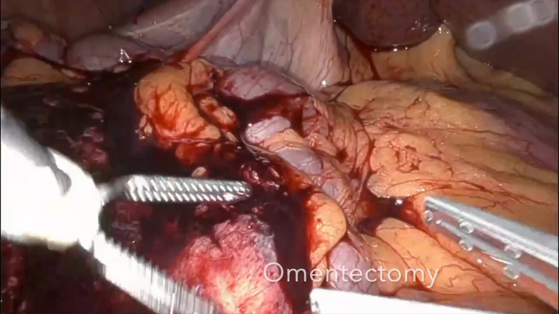
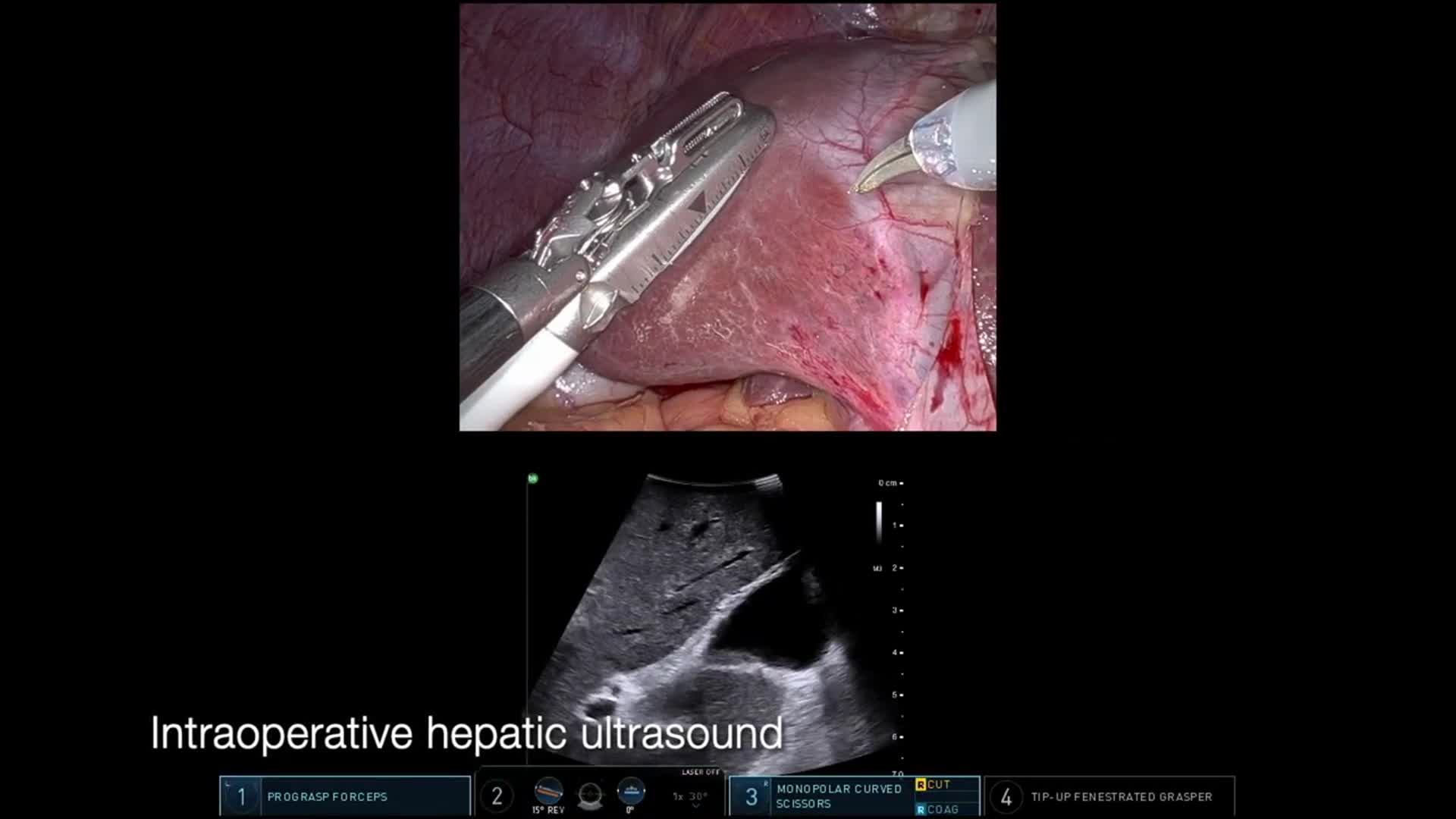
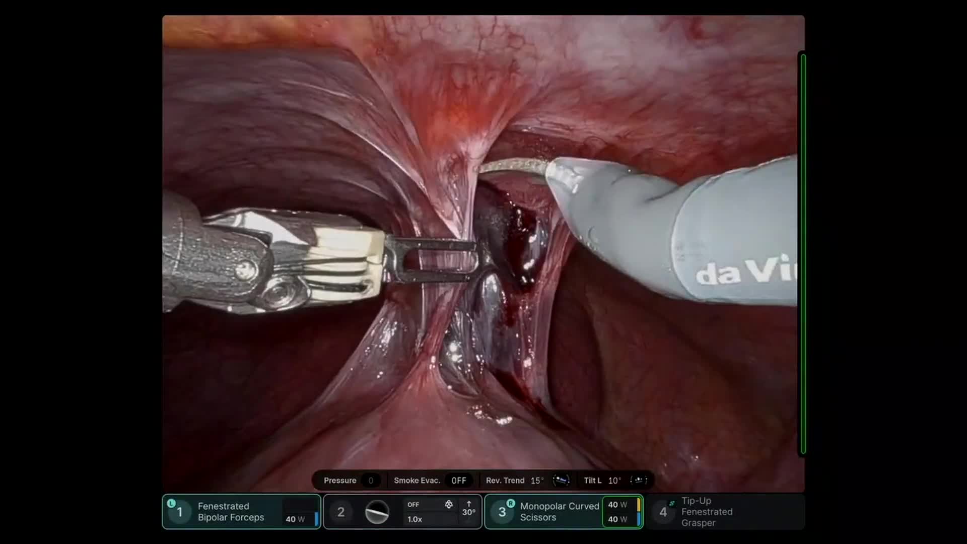
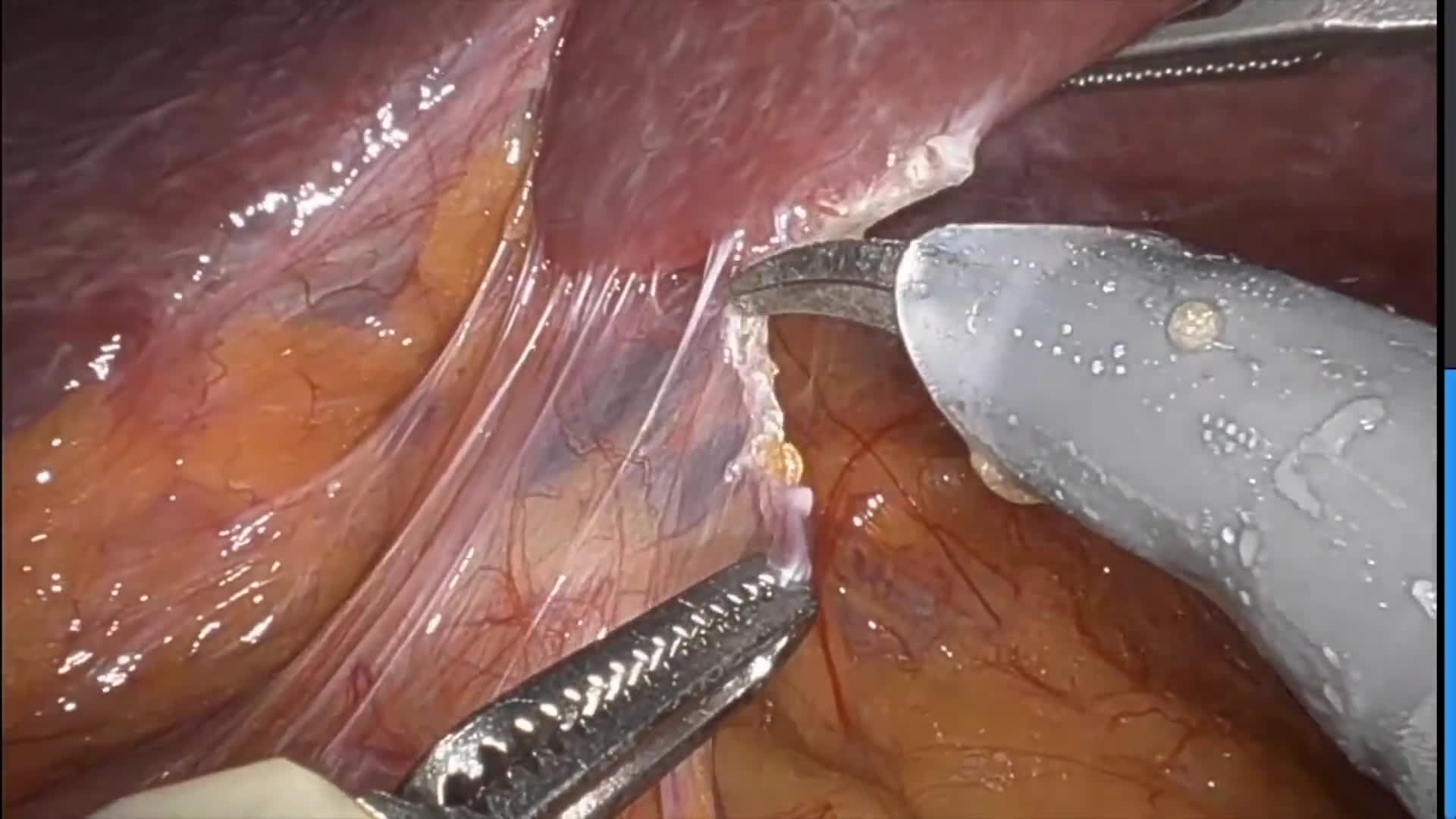
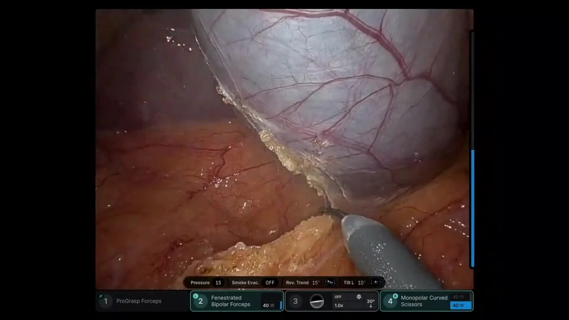
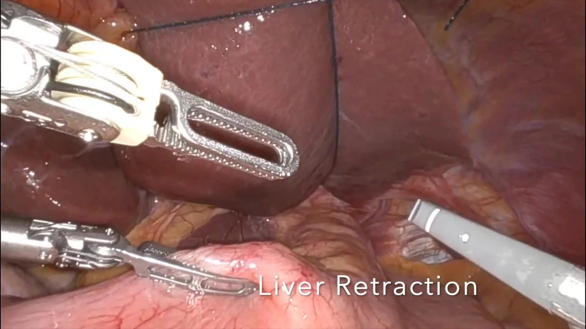
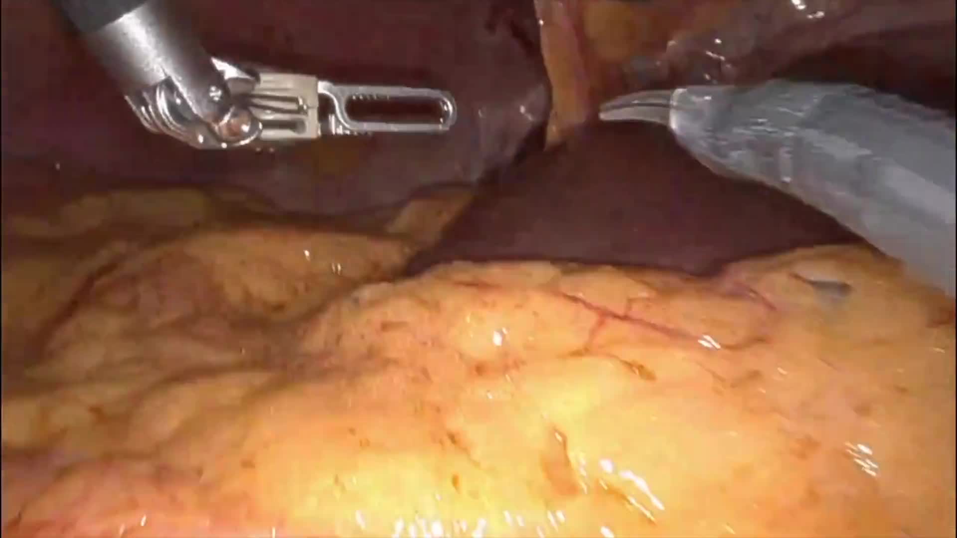
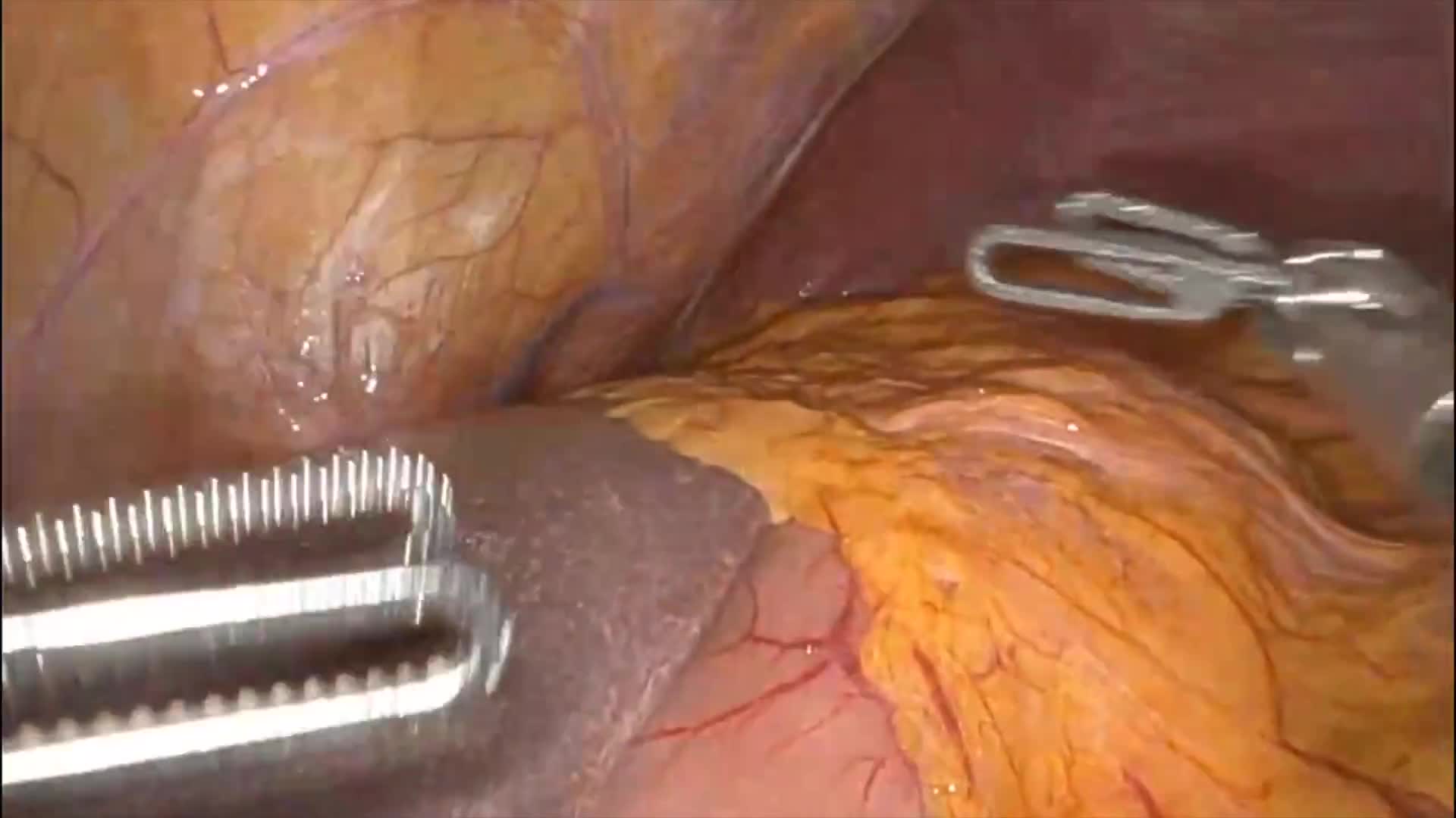
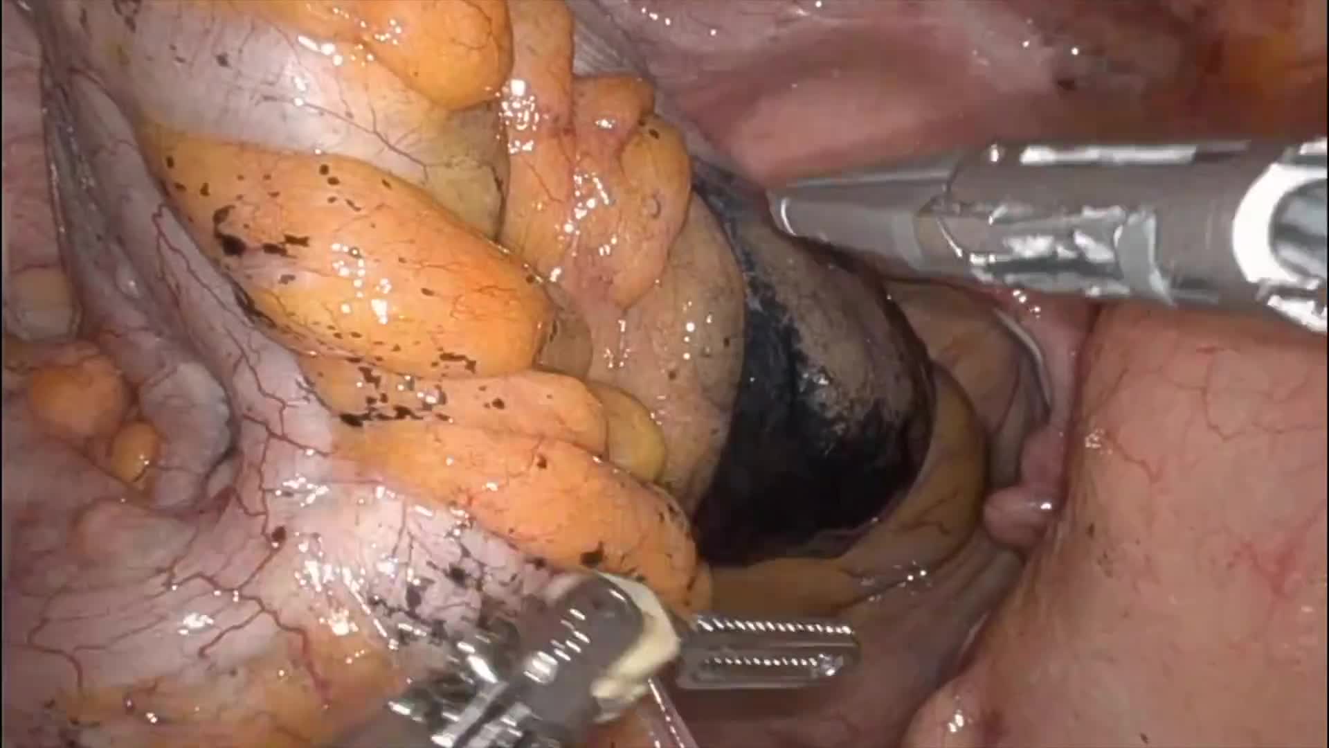
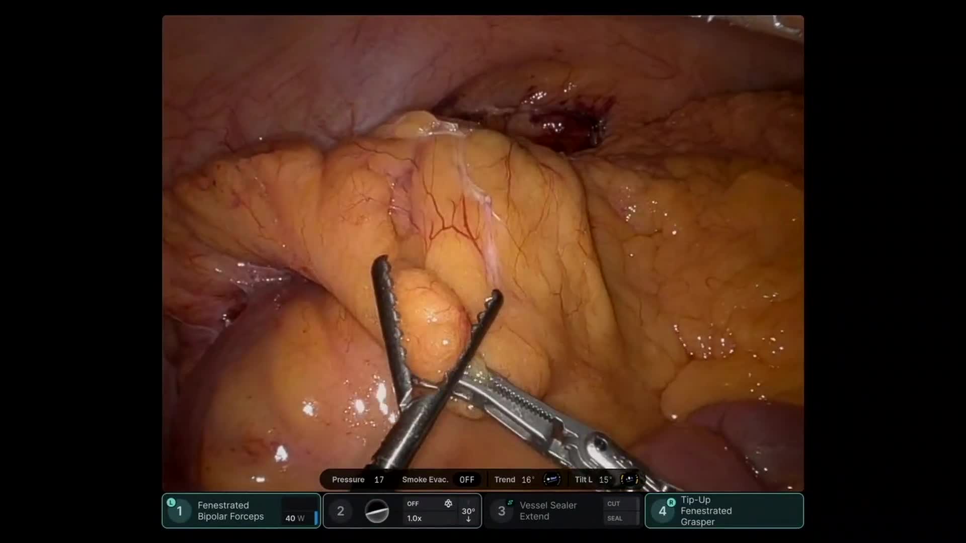
Robotic staging laparoscopy demonstrated no peritoneal metastatic disease. Intraoperative ultrasound localized a 2 cm lesion within the superior–posterior aspect of the pancreas. The lesion was carefully enucleated from the pancreatic parenchyma, and regional peripancreatic lymphadenectomy was performed.
Final pathology demonstrated a well-differentiated grade 1 pancreatic neuroendocrine tumor (Ki-67 ~1.3%). Importantly, the tumor was surrounded by pancreatic parenchyma without associated lymphoid tissue, supporting the interpretation that the lesion represented a primary pancreatic neuroendocrine tumor rather than nodal metastasis. Five additional lymph nodes were negative for malignancy.
This case illustrates the diagnostic complexity of neuroendocrine tumor staging, and demonstrates robotic techniques for suprapancreatic exposure, intraoperative ultrasound–guided localization, pancreatic tumor enucleation, and regional lymphadenectomy.